Khanna Traders, are producer and supplier of scientific equipment.
- khannachahat05@gmail.com
-
+91-8930598097
Khanna Traders, are producer and supplier of scientific equipment.
+91-8930598097


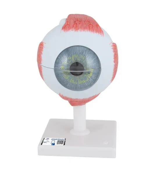
The Giant Eye replica is a great tool to teach-learn the anatomy of the eye! Removable parts of the human eye model include Lens, iris, Vitreous humour, retina
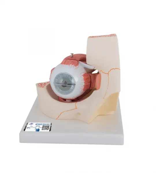
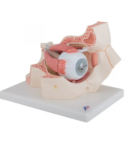
This large anatomical human eye model shows the optic nerve in its natural position in the bony orbit of the eye (floor and medial wall). At three times life size this eye model is great for anatomical demonstrations.
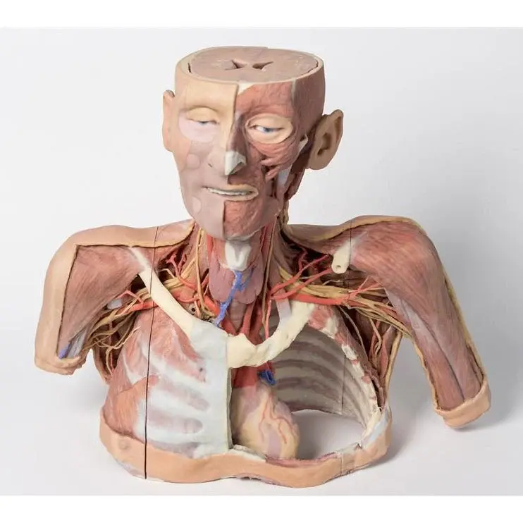
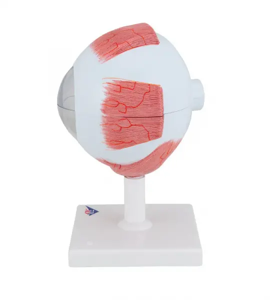
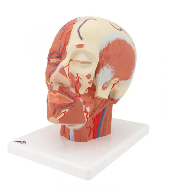
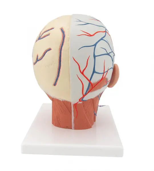
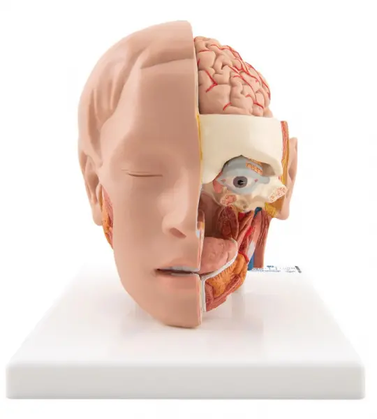
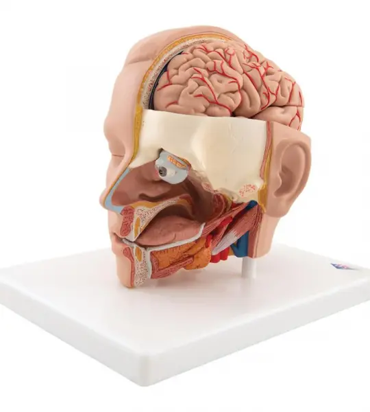
Representation of the superficial musculature with Parotid gland, Submandibular gland (right half), Deep musculature (left half).
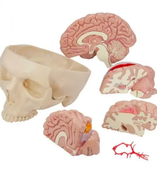
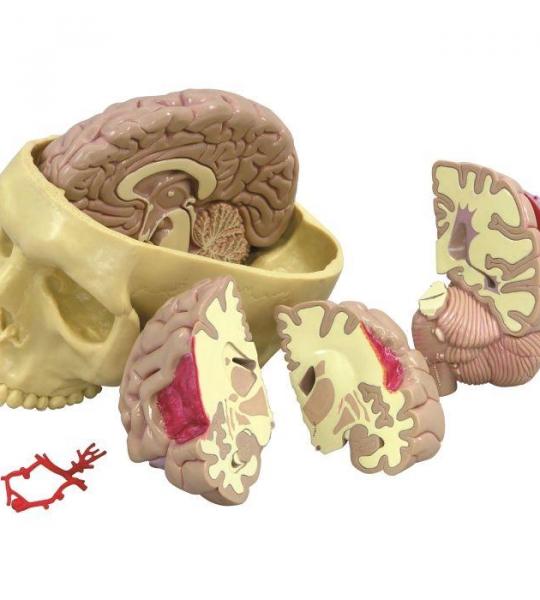
Full size segmented brain features half normal side and three-piece sectioned pathology half, as well as Circle of Willis with aneurism. The brain, which sits inside a partial skull, features the following pathologies which are also illustrated on a two-sided education card: alcoholism, Alzheimer’s, aneurism, depression related tumor, seizure related tumor, migraine, multiple sclerosis, Parkinson’s disease, stroke, and subdural hematoma.
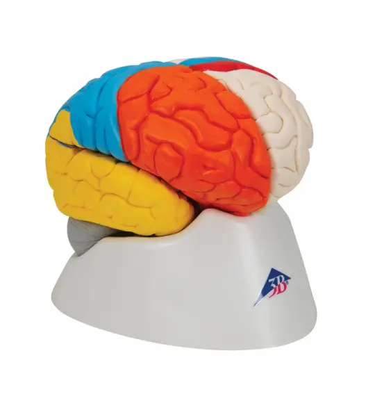
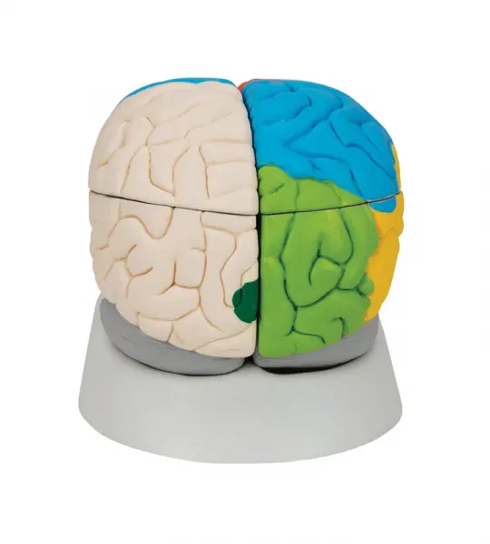
This deluxe brain is medially divided. On the right half of this brain, you will find a colored, systematic grouping and representation of the cerebral lobe.
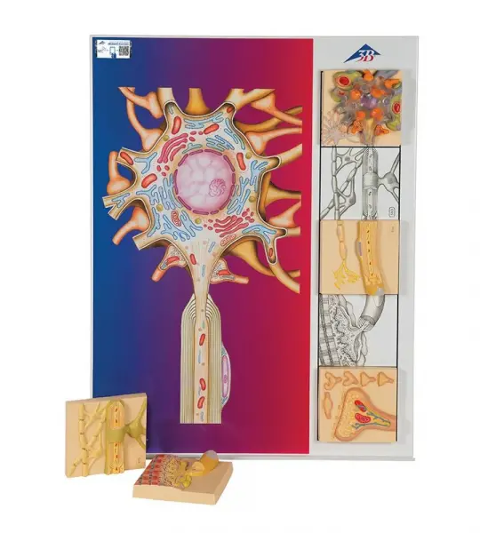
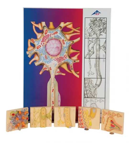
The Physiology of Nerves series displays the basic structures of the human nervous system. Each of the five sections of the nerve model shows a plastic colored relief model of the main synapse variations. All sections of the nerve physiology series can magnetically attach to the illustrated base which depicts the neural components in vivid colors. Each nerve section is also available separately.
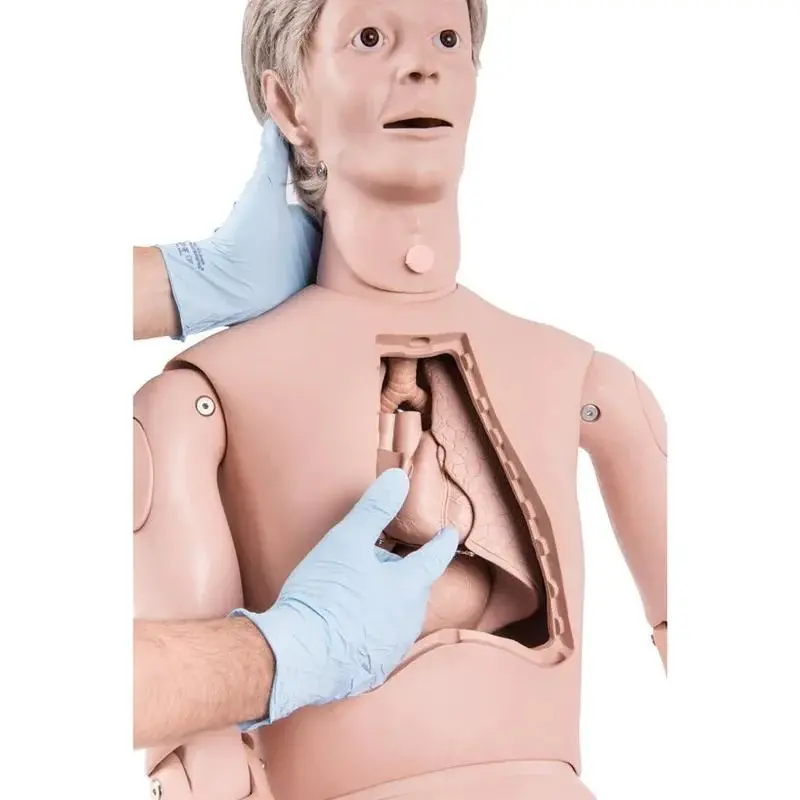
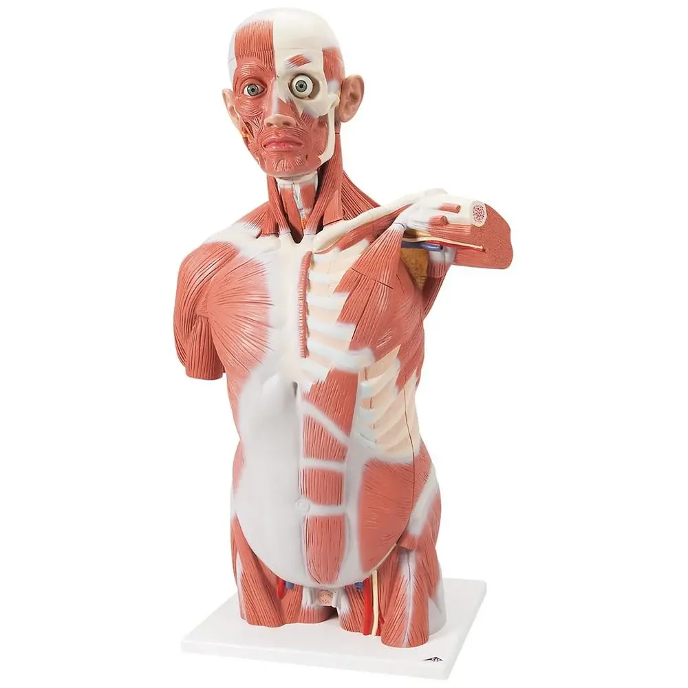
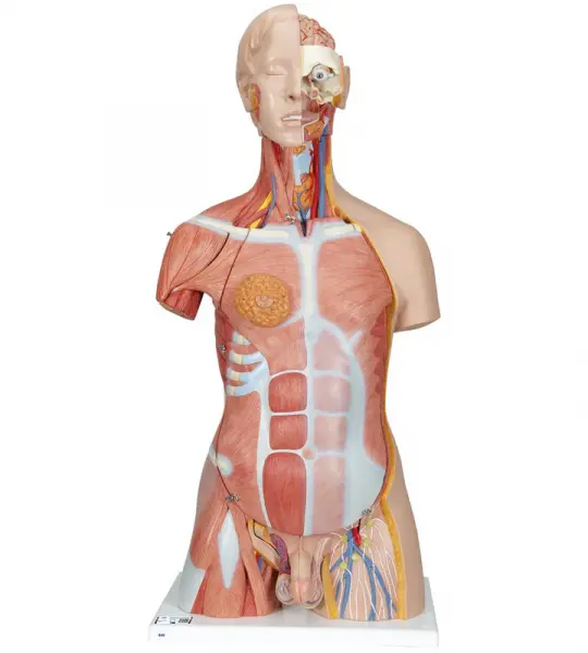
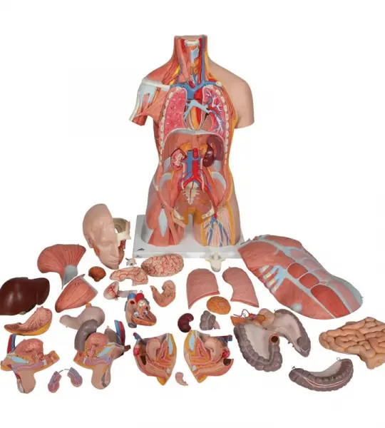
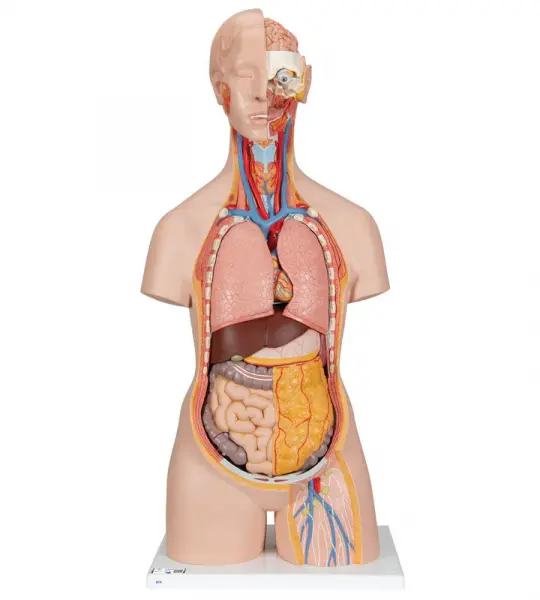
This deluxe human torso model is top notch in the field of anatomy. This unique torso depicts both the superficial and deep muscles, and the two main muscles, the deltoid and gluteus maximus can even be removed for closer studies. With this human torso model you can also study the vertebrae, the spinal cord, spinal nerves and vertebral arteries, exchange the male and female genital inserts, discover the internal structures of the brain and much more.
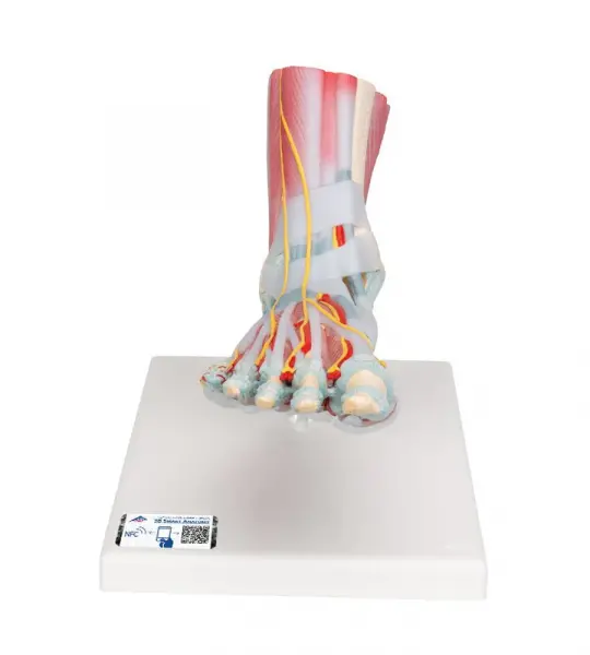
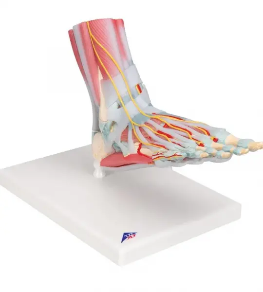
The foot skeleton features not only the bones but also the muscles, tendons, ligaments, nerves, arteries, and veins of the foot. The frontal view of the foot model features the extensor muscles of the lower leg. The tendons can be followed on their passage under the transverse and crucial crural ligaments all the way to their insertion points.


The Giant Eye replica is a great tool to teach-learn the anatomy of the eye! Removable parts of the human eye model include Lens, iris, Vitreous humour, retina
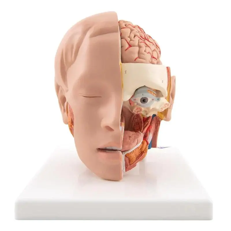
Our most detailed head model! This life-size 6-part head is mounted on a base and features a removable 4-part brain half with arteries.
This large anatomical human eye model shows the optic nerve in its natural position in the bony orbit of the eye (floor and medial wall). At three times life size this eye model is great for anatomical demonstrations.
This deluxe brain is medially divided. On the right half of this brain, you will find a colored, systematic grouping and representation of the cerebral lobe.
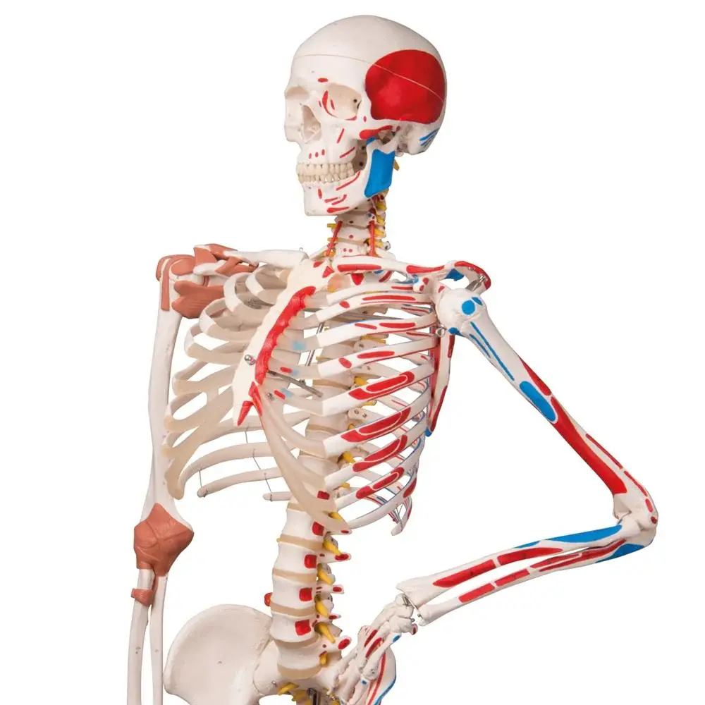
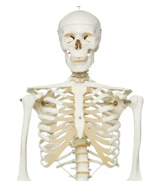
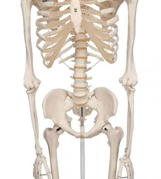
Proven Quality - even more stable! Stan, the standard model of a human skeleton, has been appreciated throughout the world for decades. Thanks to its very high quality and robust construction, it is perfect for use in hospitals, schools, universities and laboratories. So choose Stan, the original among artificial skeletons.
Representation of the superficial musculature with Parotid gland, Submandibular gland (right half), Deep musculature (left half).
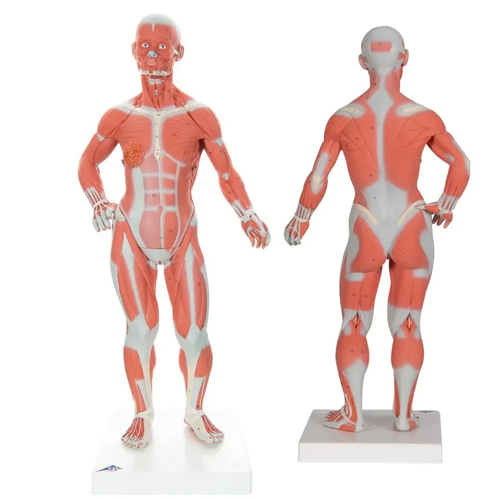
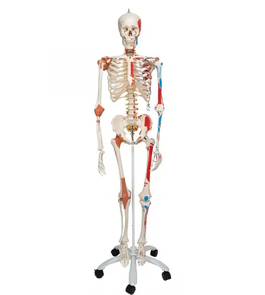
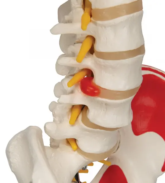
Sam offers all the advantages of a 3B Scientific® skeleton. With Sam, you can also demonstrate the movements of the skull via the head joints, and thanks to the fully flexible spine, you can adjust the model to place it in natural body postures. The unique combination of muscle origins and insertions, flexible ligaments and flexible spine with a slipped disc between the 3rd and 4th lumbar vertebrae clearly the show medical and anatomical interest of this top model's more than 600 structures. Now available on a stable metal hanging stand with 5 casters!
The foot skeleton features not only the bones but also the muscles, tendons, ligaments, nerves, arteries, and veins of the foot. The frontal view of the foot model features the extensor muscles of the lower leg. The tendons can be followed on their passage under the transverse and crucial crural ligaments all the way to their insertion points.
This deluxe human torso model is top notch in the field of anatomy. This unique torso depicts both the superficial and deep muscles, and the two main muscles, the deltoid and gluteus maximus can even be removed for closer studies. With this human torso model you can also study the vertebrae, the spinal cord, spinal nerves and vertebral arteries, exchange the male and female genital inserts, discover the internal structures of the brain and much more.
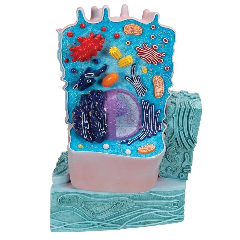
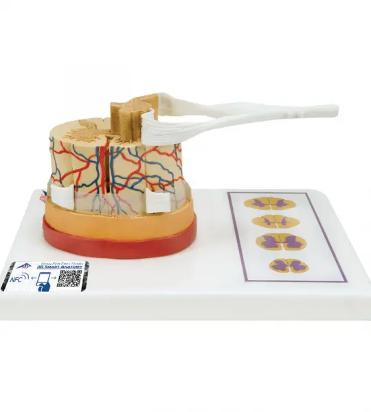
The Spinal cord model illustrates the composition of the spinal cord, magnified to a scale of about 5:1. The spinal cord is formed by a central channel surrounded by "gray matter" with an outer layer of "white matter".
The Giant Eye replica is a great tool to teach-learn the anatomy of the eye! Removable parts of the human eye model include Lens, iris, Vitreous humour, retina
This large anatomical human eye model shows the optic nerve in its natural position in the bony orbit of the eye (floor and medial wall). At three times life size this eye model is great for anatomical demonstrations.
Our most detailed head model! This life-size 6-part head is mounted on a base and features a removable 4-part brain half with arteries.
Full size segmented brain features half normal side and three-piece sectioned pathology half, as well as Circle of Willis with aneurism. The brain, which sits inside a partial skull, features the following pathologies which are also illustrated on a two-sided education card: alcoholism, Alzheimer’s, aneurism, depression related tumor, seizure related tumor, migraine, multiple sclerosis, Parkinson’s disease, stroke, and subdural hematoma.
This deluxe brain is medially divided. On the right half of this brain, you will find a colored, systematic grouping and representation of the cerebral lobe.
The Spinal cord model illustrates the composition of the spinal cord, magnified to a scale of about 5:1. The spinal cord is formed by a central channel surrounded by "gray matter" with an outer layer of "white matter".
Free Delivery
Worldwide
Money Returns
30 Days money return
27/4 Support
Customer Support
Payment Security
Safe Payment